Fluoroscopy Can Best Be Described as
Fluoroscopy is a study of moving body structures--similar to an X-ray movie A continuous X-ray beam is passed through the body part being examined. Fluoroscopy is an imaging technique that uses X-rays to obtain real-time moving images of the interior of an object.
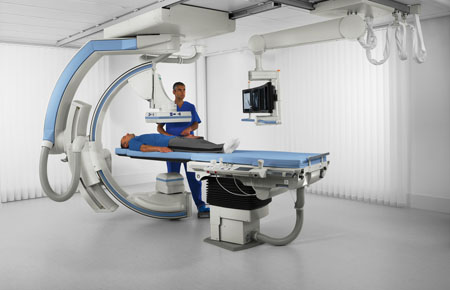
Penunjang Medis C Arm Radiografi Dan Fluoroscopy 3d Rumah Sakit Akademik Ugm
Fluoroscopy can identify plantar plate tear.

. Welcome to GIGU Fluoroscopy Introduction. For purposes of instructing students and residents we have divided pulmonary fluoroscopy into five phases. This is clinically significant because it requires timely surgical repair to prevent complications such as loss of push-off strength in gait and cock-up toe.
Medical imaging can provide life-saving information so the benefits of the procedure generally far outweigh the risks from. X-rays are a form of ionizing radiation. While many answers can be provided through the use of cross-.
The chest is included beucase the kidneys are located under the lowest ribs. Fluoroscopy is an imaging technique used by medical professionals to visualize internal organs while they are in motion. In its primary application of medical imaging a fluoroscope allows a physician to see the internal structure and function of a patient so that the pumping action of the heart or the motion of swallowing for example can be watched.
Early fluoroscopes were simple boxes made of cardboard that were open at one end the. Fluoroscopy is a medical imaging test that uses X-rays to obtain still and moving images of structures and processes inside the body often with use of a contrast agent. As with the photospot camera we can calculate the effective resolution of the film at the face of the image intensifier.
Fluoroscopy can be traced back to 1895 when Wilhelm Röntgen noticed a barium platinocyanide screen fluorescing due to exposure to what he would later define as x-rays. Radiation exposure in pediatric fluoroscopy can be reduced to values well below the exposure settings that are typically found on unoptimized fluoroscopes. Fluoroscopy can diagnose or aid in diagnosis of many conditions.
A continuous X-ray beam is passed through the body part and sent to a video monitor. This result is significantly better than the 5 lpmm offered by the best intensifiers. Usually best seen in the left oblique view Fig.
If the film resolves 200 lpmm the effective resolution at the intensifier input would be 200 18230 or 16 lpmm. Individual hospital coding guidelines determine whether the fluoroscopy is separately coded. This is useful for both.
Fluoroscopy can be used for diagnosing finding out the cause of a health problem such as heart or intestinal disease. If an X-ray is a still picture fluoroscopy is like a movie. Fluoroscopic procedures expose patients to a greater amount of radiation than other medical imaging procedures such as x-rays.
X ray of the chest and abdomen while the patient is lying on the x ray table. Fluoroscopy as an imaging tool enables physicians to look at many body systems including the skeletal digestive urinary. Fluoroscopy is a technique that provides real-time X-ray imaging that is especially useful for guiding a variety of diagnostic and interventional procedures.
The beam is transmitted to a TV-like monitor so that the body part and its motion can be seen in detail. Left to right B. Fluoroscopy is a type of imaging procedure that uses several pulses of an X-ray beam to take real-time footage of tissues inside your body.
Right to left C. The fluoroscopy is included in the bronchoscopy and no code is assigned for it. The X-rays make the tracer glow or fluoresce showing the structure and real-time function of the organ or system on a screen.
Moving images of the inside of your body. Fluoroscopy is a type of imaging tool. Fluoroscopy is a medical procedure that makes a real-time video of the movements inside a part of the body by passing x-rays through the body over a period of time.
In some cases fluoroscopic images may be stored as part of the patient examination. It refers to using X-rays from a CT scanner to bounce off a mildly radioactive tracer whether swallowed administered as an enema or injected into veins. This may be the only clue to a correct pre-operative diagnosis of pulmonary.
Fluoroscopy can be accomplished within a few minutes if the examiner applies a well-organized approach. The body part and its motion can then be seen in detail. Appointment Center 247 2164457050.
The aim of the Fluoroscopy Service at UF is to provide complete and accurate studies performed in an expeditious manner with attempts to minimize patient discomfort and delay while assisting referring physicians in answering clinical conundrums. Pulsed fluoroscopy was able to lower radiation dose to less than 10 of continuous fluoroscopy while still maintaining acceptable phantom image quality. Left to right B.
Net movement of glucose across the membrane can best be described as A. The images are projected onto a monitor very similar to a television screen. -the motion of organs can be seen with continuous x rays.
Its much like an X-ray movie It is often done while a contrast dye moves through the part of the body being examined. Right to left C. Fluoroscopy-guided catheter angiography is an interventional procedure that uses percutaneous access of arteries with needles and catheters to inject contrast for vessel opacification1 This procedure may be diagnostic or therapeutic.
Fluoroscopy is frequently used to assist in a wide variety of medical diagnostic and therapeutic procedures both within and outside of. It looks at moving body structures. Percutaneous transthoracic aspiration biopsi.
Movement of glucose across the membrane can best be described as A. The fluoroscopy guidance code is assigned as a secondary procedure code with the bronchoscopy code. Since the procedure is done in real-time the movement of different organs and structures can be visualized.
The first fluoroscopes were invented several months after Röntgens discovery of x-rays. Process of using an instrument to examine x-rays transformed into fluorescence. The fluoroscopy guidance code is assigned as the first procedure code.
Its important to discuss the necessity of the procedure with your medical professional. The purpose of this study was to evaluate the feasibility of using an open-configuration magnetic resonance MR imaging system with MR fluoroscopic guidance to perform percutaneous transthoracic fine-needle aspiration biopsy in patients with lung masses. Healthcare providers use fluoroscopy to help monitor and diagnose certain conditions and as imaging guidance for certain procedures.

R F Radiography Fluoroscopy Can Be Described As The Initial Stage Of Human Imaging Studies Over The Years Th Hospital Interior Room Interior Operating Room

0 Response to "Fluoroscopy Can Best Be Described as"
Post a Comment